DiR Iodide (DiIC18(7)) 细胞膜深红色荧光探针
货号: JP4005-5MG 品牌: Jinpan 标签: 细胞生物学研究
描述
DiR Iodide (DiIC18(7)) 细胞膜深红色荧光探针
温馨提示:见细胞膜荧光探针专题,选择您想要的最适膜标记探针。
搜索关键词:
细胞膜荧光探针,DiI,DiO,DiD,DiR,DiA,CM-DiI,神经示踪,传统细胞膜荧光探针
订购信息:
| 产品名称 | 产品编号 | 规格 | 价格(元) |
| DiR Iodide (DiIC18(7)) 细胞膜深红色荧光探针 | JP4005-5MG | 5mg | 398.00 |
| DiR Iodide (DiIC18(7)) 细胞膜深红色荧光探针 | JP4005-25MG | 25mg | 1188.00 |
产品描述:
DiI, DiO, DiD和DiR作为一类长链的亲脂性二烷基碳菁类染料(Dialkylcarbocyanines)荧光染料家族,用于标记细胞膜以及其他脂溶性生物结构。作为一类环境敏感型荧光染料,当它们与膜结合或者与亲脂性生物分子(例如蛋白质,虽然在水中其荧光强度很弱)结合时,其荧光强度显著增强。一旦进入细胞后,它们在细胞内质膜中逐步扩散,于最佳浓度条件下可将整个细胞均匀染色。这些染料具很高的淬灭系数,偏光依赖性和很短的激发寿命。
四种染料呈现不同的荧光颜色,DiI(橙色荧光)、DiO(绿色荧光)、DiD(红色荧光)、DiR(深红色荧光),为活细胞多色彩荧光成像分析和流式细胞术提供了一种便捷的工具。DiO和DiI分别用标准FITC和TRITC滤光器检测,可结合使用。DiD可被633 nm氦-氖(He-Ne)激光激发,具有比DiI更长的激发和发射光波长,特别适合用于标记具本底荧光的细胞和组织。而,DiR在体内活体成像或者示踪中意义非凡,因其所发射的红外光可以高效地穿过细胞和组织,并且在红外光范围内,其本底荧光水平很低。
本品以DiR的碘盐形式提供,英文名:1,1-dioctadecyl-3,3,3,3-tetramethylindotricarbocyaine iodide,纯度≥95%,适用于荧光检测研究。
基本特性:
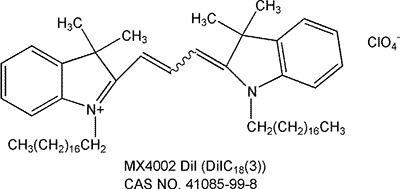
1)同义名:DiR Iodide; DiIC18(7);1,1-dioctadecyl-3,3,3,3-tetramethylindotricarbocyaine iodide;
2)推荐滤光器:Omega-XF112, Chroma-41009
3)分子式:C63H101IN2
4)分子量:1013.39g/mol
5)外观:蓝色至深蓝色固体
6)纯度:≥95%(HPLC)
7)Ex/Em:750/780 nm(甲醇)
8)溶解性:溶于DMF,DJPO,甲醇
9)化学结构图: 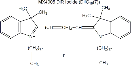

保存与运输方法
保存:-20ºC避光干燥保存,有效期二年。
运输:冰袋运输。
应用示例(来自文献)
文献来源:Eisenblätter M et al. In Vivo Optical Imaging of Cellular Inflammatory Response in Granuloma Formation Using Fluorescence-Labeled Macrophages. J Nucl Med. 2009 Oct;50(10):1676-82.
标记方法(Labeling Protocol):脂质示踪剂DiR(1,1-dioctadecyl-3,3,3,3-tetramethylindotricarbocyanine iodide),最大激发或者发射波长位于近红外区域(Ex/Em:750/782nm),溶于乙醇(0.98mM)。预实验中用梯度浓度的DiR(0.98~197nM)来确定标记效率。后续实验中,单核细胞(1 × 105/mL培养基)加入2ul DiR标记液(终浓度为19.7uM)孵育5min,PBS清洗3次后,重悬于细胞培养液中。流式细胞仪检测标记效率。
Cutaneous Granuloma (CG) Model(皮肤肉芽肿体内模型):在小鼠侧区皮下注射1ml 聚丙烯酰胺凝胶(PAG)诱导皮肤肉芽肿形成。为了诱导炎症,向PAG内加入脂多糖LPS(10ug LPS/ml)。在凝胶移植前24h静脉注射DiR标记的巨噬细胞或对照巨噬细胞。使用FRI和FMT在注射后每日观察小鼠,持续7天。
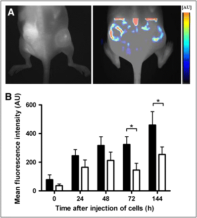
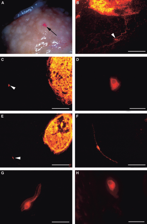
FIG 1. FMT of mice showed distribution of Mϕs inside pellets. (A) FMT was performed 72 h after injection of labeled Mϕs (5 × 10⁶) to resolve 3-dimensional distribution of Mϕs in target tissue (left, NIRF image; right, color-encoded FMT). FMT confirmed FRI results and showed that Mϕs distributed mainly in periphery of pellets. (B) Fluorescence quantification by FMT showed approximately 2.7 times higher signal in lipopolysaccharide-containing pellets (▪) than in controls (□).
文献来源:Jiang H et al. Liver-targeted liposomes for codelivery of curcumin and combretastatin A4 phosphate: preparation, characterization, and antitumor effects. Int J Nanomedicine. 2019 Mar 8;14:1789-1804.
DiR标记GA LPs的制备(Preparation of DiR-loaded GA LPs):First, 120 mg l-α-phosphatidylcholine, 40 mg Chol, 20 mg DSPE-PEG2,000-GA, and 0.2 mg DiR were mixed in 5 mL Chol at a mass ratio of 600:200:100:1 in an eggplant-shaped flask, dried until a thin-lipid film had formed on the rotary evaporator under reduced pressure, and heated in a 35°C water-bath. The film was hydrated with 5 mL PBS, followed by sonication. The obtained DiR-GA LPs were filtered in a 220 nm filter device, and the final concentration of DiR was 40 µg/mL.
体内实时近外荧光成像(In vivo real-time near-infrared fluorescence imaging): NIRF was used to observe the biodistribution of GA LPs formulation in vivo, and DiR-GA LPs (40 µg/mL) were prepared to monitor the fate of LPs. When tumors were 100 mm3, DiR-GA LPs were injected into tail veins of mice. Free DiR was used as control. Precisely 10 mg DiR iodide in a 10 mL volumetric bottle was dissolved in methanol at a constant volume and prepared into a 1 mg/mL drug-reserve solution, which was stored at 4°C in the refrigerator for later use. DiR iodide reserve solution diluted with normal saline to the concentration of 40 µg/mL. Mice were anesthetized with 10% chloral hydrate and real-time NIRF images obtained with excitation and emission at 745 and 835 nm, respectively.44 Results were analyzed using the Living Image 3.1 software.
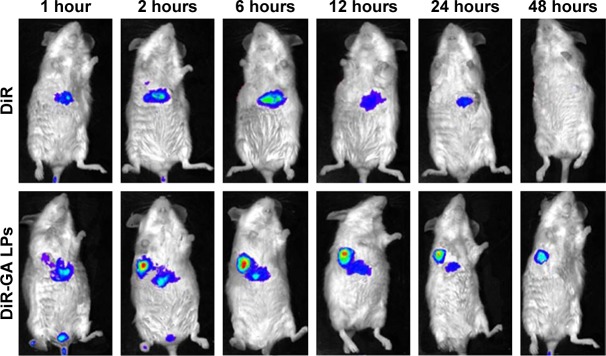
FIG2. NIRF images of H22 tumor-bearing mice after injection of free DiR and DiR-GA LPs. Results showed that NIRF signals of the free-DiR group were lower than the DiR-GA LPs group from 1 to 48 hours. No fluorescence signals in the tumor regions were detected for the free-DiR group, except at 2 hours. By contrast, DiR-GA LPs increased in accumulation in tumor regions and signals were sustained at 48 hours after injection. The GA LP drug loading system increased the accumulation of DiR in tumors.
注意事项
1)荧光染料均存在淬灭问题,请尽量注意避光,以减缓荧光淬灭。
2)为了您的安全和健康,请穿实验服并戴一次性手套操作。
使用方法
【注意】以下使用方法仅用作参考,可根据具体的实验条件做出调整。
1. 染色液制备
1)储存液制备:用DJPO或乙醇配置浓度1~5 mM的储存液。例如,取25mg DiR(Mw:1013.39g/mol)溶于4.93ml无水DJPO中,充分溶解,即得到5mM的储存液。【注意】未使用的储存液分装储存在-20℃,避免反复冻融,且要避光保存。
2)工作液制备:用合适的缓冲液(如:无血清培养基,HBSS或PBS)稀释储存液,调整到1~5 μM的工作浓度。【注意】工作液最终浓度需要根据不同细胞系和实验体系来优化。建议从推荐浓度开始,以10倍范围为区间进行最优浓度的摸索。
2. 悬浮细胞染色
1)加入适当体积的染色工作液重悬细胞,使其密度为1×10⁶/mL。
2)37℃孵育细胞2~20min,不同的细胞最佳培养时间不同。可以20min作为起始孵育时间,之后优化以保证得到均一化的标记结果。
3)孵育结束,按1000~1500 rpm离心5min。
4)去除上清液,之后轻柔加入37℃预热的生长培养液重悬细胞。
5)再重复3),4)步骤两次。
3. 贴壁细胞染色
1)将贴壁细胞培养于无菌盖玻片上。
2)从培养基中移走盖玻片,滤掉过量培养液,将盖玻片放在潮湿的小室内。
3)在盖玻片的一角加入100μL的染色工作液,轻轻晃动使染料均匀覆盖所有细胞。
4)37℃孵育细胞2~20min,不同的细胞最佳培养时间不同。可以20min作为起始孵育时间,之后优化以保证得到均一化的标记结果。
5)吸掉染色工作液,用培养液洗盖玻片2~3次,每次用预温的培养基覆盖所有细胞,孵育5~10min,然后吸走培养基。
4. 显微镜检测
1)DiD,DiO,DiI,DiR和DiS滤光器的选择参见下表:
| 货号 | 荧光探针 | 最大激发/最大发射波长
(Ex/Em) |
滤光片编号 | |
| Omeaga公司 | Chroma公司 | |||
| JP4002-10MG | DiI | 549/565nm | XF108, XF32 | 41002, 31002 |
| JP4001-25MG | DiO | 484/501nm | XF100,XF2341001, 31001 | 41001, 31001 |
| JP4004-25MG | DiD | 644/665nm | XF110, XF47 | 41008, 31023 |
| JP4005-25MG | DiR | 750/780nm | XF112 | 41009 |
2)多色染料的同时检测,滤光器按照以下设定:
a) DiI和DiO=Omega XF52,Chroma 51004;
b) DiI和DiD=Omega XF92,Chroma 51007;
c) DiI,DiO和DiD=Omega XF93,Chroma 61005;
5. 流式细胞仪检测
经DiO,DiI,DiD和DiR染色的细胞分别用流式细胞仪的FL1,FL2,FL3或FL4通道检测。
规格信息
| 品牌: |
Jinpan |
|---|---|
| CAS: |
N/A |
| 规格: |
5mg |
| 货期: |
咨询客服 |