8-羟基-(脱氧)鸟嘌呤定量ELISA分析——DNA/RNA损伤分析

活性氧(ROS)自由基可以直接攻击生物大分子DNA/RNA诱发DNA/RNA氧化损伤。DNA分子的损伤类型有多种。UV照射后DNA分子上的两个相邻的胸腺嘧啶 (T)或胞嘧啶(C)之间可以共价键连结形成环丁酰环,这种环式结构称为二聚体。DNA 分子还可以发生个别碱基或核苷酸的变化。例如碱基结构类似物5-溴尿嘧啶等可以取代个别碱基等。

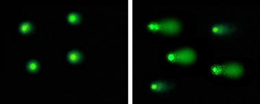
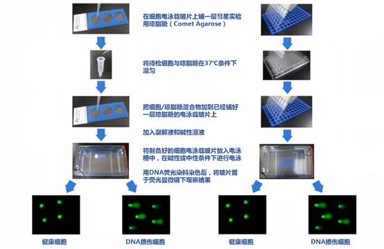
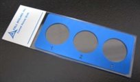
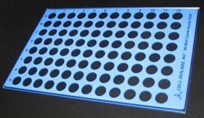
针对DNA/RNA损伤的检测,上海金畔生物科技有限公司向您推荐Cell BioLabs完整的DNA/RNA氧化损伤检测方案,其OxiSelect™ Oxidative DNA / RNA Damage ELISA Kits(8-OHdG or 8-OHG Quantitation)提供了快速高效检测核酸中8-OHG或8-OHdG含量的实验方案;OxiSelect™ Comet Assays (Single Cell Gel Electrophoresis)则为您提供多种选择的彗星分析产品;针对DNA损伤中的AP位点检测,Cell Biolabs的OxiSelect™ Oxidative DNA Damage Quantitation Kit (AP Sites)则使用经典的ARP检测方案;DNA双链断裂分析为您提供了快捷的DSB分析试剂。

在各种氧化损伤中,以鸟嘌呤8位碳原子氧化后形成8-羟基-[脱氧]鸟嘌呤(8-OHdG/8-OHG)最为常见。还有一些嘌呤或者嘧啶碱基直接脱去的反应,这样也就形成了核酸中无嘌呤(apurinic)和脱嘧啶(apyrimidinic)位点,统简称AP位点。除了上述氧化损伤外,DNA双链断裂(DSBs)是细胞内多种类型的DNA损伤中最危险、最8-羟基-(脱氧)鸟嘌呤定量ELISA分析——DNA/RNA损伤分析严重的一种。上海金畔生物科技有限公司向您推荐Cell BioLabs完整的DNA/RNA损伤检测方案,其产品覆盖上述最重要的DNA/RNA损伤分析产品,为您提供最优的检测方案。

其中OxiSelect™ Oxidative DNA / RNA Damage ELISA Kits(8-OHdG or 8-OHG Quantitation)提供了快速高效检测核酸中8-OHG或8-OHdG含量的实验方案。在各种氧化损伤中,以鸟嘌呤8位碳原子氧化后形成8-羟基-脱氧鸟嘌呤(8-OHdG)最为常见8-羟基-脱氧鸟嘌呤(8-OHdG)因此也成为DNA损伤最普遍的标志物。作为DNA损伤的副产物,当有化学致癌物刺激时,会产生更多的8-OHdG。在生物体内核酸外切酶修复损伤DNA时,形成的8-羟基-脱氧鸟嘌呤会被分泌出去而不会进行尿液中进行进一步的代谢。同样的情况也在RNA中发生,只不过是由鸟嘌呤8位碳原子氧化后形成8-羟基-鸟嘌呤(8-OHG),其也被作为RNA损伤的重要检测指标。

Cell Biolabs的OxiSelect™ Oxidative DNA / RNA Damage ELISA Kits试剂盒采用竞争性ELISA方法定量8-OHdG/8-OHG。待测样品和8-OHdG/8-OHG标准样分别加入8-OHdG/8-OHG-BSA联结的EIA板内,孵育一段时间后,加入anti-8-OHdG/anti-8-OHG单抗,然后加入HRP联结的二抗,通过与8-OHdG/8-OHG标准曲线比对获得最终数据。该试剂盒检测灵敏,能有效检测样品中100pg/ml的8-OHdG或300pg/ml的8-OHG含量,每个试剂盒能完成至少96次分析。

产品名称 货号 产品说明(点击查看说明书)
OxiSelect™ Oxidative DNA Damage ELISA Kit (8-OHdG Quantitation) STA-320 ELISA比色法检测,96次分析
STA-320-5 ELISA比色法检测,5×96次分析
OxiSelect™ Oxidative RNA Damage ELISA Kit (8-OHG Quantitation) STA-325 ELISA比色法检测,96次分析
STA-325-5 ELISA比色法检测,5×96次分析

上海金畔生物科技有限公司向您推荐Cell BioLabs完整的DNA/RNA损伤检测方案,其产品覆盖上述最重要的DNA/RNA损伤分析产品,为您提供最优的检测方案。除了传统的8-OHdG/8-OHG定量、AP位点分析等,还有基于单细胞水平的彗星分析和DNA双链断裂分析。除了DNA/RNA损伤,还包括脂质过氧化过程中壬烯(HNE)、丙二醛(MDA)以及8-异前列腺素F2a(8-Isoprostane)ELISA分析检测试剂盒;对于蛋白质的羰基化、硝基化以及终末氧化蛋白产物分析等蛋白氧化损伤检测方案;针对活性氧基团和抗氧化剂的活力检测,也由多种试剂和试剂盒供您选择。如果您对以上产品感兴趣,请致电021-50837765到上海金畔生物科技有限公司垂询氧化应激及损伤相关的实验解决方案,或索取最新的Cell BioLabs产品资料。

Cell Biolabs彗星实验试剂盒

由于环境因素和细胞内的正常代谢过程造成的DNA损伤,每个细胞每天都会发生1,000到1,000,000个。虽然这些只占人类基因组约60亿个碱基中的一小部分,但如果关键基因损伤未及时修复,可能会阻碍细胞的正常生理功能,进而增加癌变可能。彗星实验,或称单细胞凝胶电泳(Single cell gel electrophoresis,SCGE),是一种测量单个细胞DNA损伤的常用技术。其原理很简单,即在电泳场中,将受损细胞DNA(包含片段和链断裂)与完整的DNA分离,通过显微镜可观察到损伤细胞呈现出典型的彗星状尾巴,然后通过测量计算彗尾大小对比出细胞DNA损伤的程度。因为彗星实验的特点,该方法几乎被用来评估任何类型的真核细胞的 DNA 修复能力,包括双、单链断裂的不同的 DNA 损伤情况。是一种能快速、大通量检测真核细胞DNA损伤进而判别遗传毒性的技术。

Cell Biolabs彗星实验试剂盒

彗星实验结果图

彗星实验原理虽简单,但操作繁琐,需要丰富的实验经验和技巧,尤其常常出现的“脱胶”问题,困扰了许多科研人员。除此之外,有时为了跑出完美的“彗星”图案放在paper里,还需要重复做许多次实验,费时费力。为了解决上述问题,我们推荐CellBiolabs的OxiSelectTMComet Assay Kit即彗星实验试剂盒来检测细胞的DNA损伤。该试剂盒不仅能让彗星实验化繁为简,还有两种不同规格(3孔和96孔)的细胞电泳凝胶板供选择,让少量样本和大量样本的DNA损伤检测通通轻松hold住。用该试剂盒做彗星实验流程如下图。

Cell Biolabs彗星实验试剂盒

除了操作简便,OxiSelectTMComet Assay Kit还有以下优点:

1)      适用于各种DNA损伤检测,是一款非常好用的DNA损伤检测筛选工具;

2)      试剂盒中的载玻片经过特殊处理以粘附低熔点琼脂糖,避免“脱胶”问题出现;

3)      采用特殊的DNA荧光染料,能有效降低背景干扰,更加方便读取实验结果。


彗星实验试剂盒信息:

品名

货号

规格

说明

OxiSelectTM Comet Assay Kit (3-Well Slides)

STA-350

15 assays

试剂盒内有5张3孔载玻片和彗星实验所需的低熔点琼脂糖、裂解液及DNA荧光染料等,共可检测15个样品。

OxiSelectTM Comet Assay Kit (3-Well Slides)

STA-351

75 assays

试剂盒内有25张3孔载玻片和彗星实验所需的低熔点琼脂糖、裂解液及DNA荧光染料等,共可检测75个样品。

OxiSelectTM Comet Assay Kit (96-Well Slides)

STA-355

96 assays

试剂盒内有1张96孔载玻片和彗星实验所需的低熔点琼脂糖、裂解液及DNA荧光染料等,共可检测96个样品。

为了满足客户更多样的实验需求,彗星实验试剂盒内特殊处理电泳载玻片还可以单独购买,详情如下:

品名

货号

规格

产品图片

Comet Assay Slides, 3-Well

STA-352

5 slides

Cell Biolabs彗星实验试剂盒

STA-353

25 slides

Comet Assay Slides, 96-Well

STA-356

1 slides

Cell Biolabs彗星实验试剂盒

STA-356-5

5 slides

产品部分发表文献:

  • Wang, T. et al. (2021). The effects of glucose-6-phosphate dehydrogenase deficiency on benzene-induced hematotoxicity in mice. Ecotoxicol Environ Saf. 226:112803. doi: 10.1016/j.ecoenv.2021.112803.
  • Ciminera, A.K. et al. (2021). Elevated glucose increases genomic instability by inhibiting nucleotide excision repair. Life Sci Alliance. 4(10):e202101159. doi: 10.26508/lsa.202101159.
  • Hudita, A. et al. (2021). Bioinspired silk fibroin nano-delivery systems protect against 5-FU induced gastrointestinal mucositis in a mouse model and display antitumor effects on HT-29 colorectal cancer cells in vitro. Nanotoxicology. doi: 10.1080/17435390.2021.1943032.
  • Hung, S.Y. et al. (2021). Bavachinin Induces G2/M Cell Cycle Arrest and Apoptosis via the ATM/ATR Signaling Pathway in Human Small Cell Lung Cancer and Shows an Antitumor Effect in the Xenograft Model. J Agric Food Chem. doi: 10.1021/acs.jafc.1c01657.
  • Cho, K. et al. Suppressor of cytokine signaling 2 is induced in Huntington’s disease and involved in autophagy. Biochem Biophys Res Commun. 559:21-27. doi: 10.1016/j.bbrc.2021.04.089.
  • Cho, D.H. et al. (2021). Far-infrared irradiation inhibits breast cancer cell proliferation independently of DNA damage through increased nuclear Ca2+/calmodulin binding modulated-activation of checkpoint kinase 2. J Photochem Photobiol B. doi: 10.1016/j.jphotobiol.2021.112188.
  • Li, J. et al. (2021). Melatonin ameliorates cypermethrin-induced impairments by regulating oxidative stress, DNA damage and apoptosis in porcine Sertoli cells. Theriogenology. 167:67-76. doi: 10.1016/j.theriogenology.2021.03.011.
  • Li, M.Z. et al. (2021). Discovery of MTR-106 as a highly potent G-quadruplex stabilizer for treating BRCA-deficient cancers. Invest New Drugs. doi: 10.1007/s10637-021-01096-4.
  • Jeske, R. et al. (2021). Agitation in a Microcarrier-based Spinner Flask Bioreactor Modulates Homeostasis of Human Mesenchymal Stem Cells. Biochem Eng J. doi: 10.1016/j.bej.2021.107947.
  • Zhou, W. et al. (2021). Fine polystyrene microplastics render immune responses more vulnerable to two veterinary antibiotics in a bivalve species. Mar Pollut Bull. 164:111995. doi: 10.1016/j.marpolbul.2021.111995.
  • Park, K. et al. (2021). Aicardi-Goutières syndrome-associated gene SAMHD1 preserves genome integrity by preventing R-loop formation at transcription–replication conflict regions. PLoS Genet. 17(4): e1009523. doi: 10.1371/journal.pgen.1009523.
  • Ramos, H. et al. (2021). A selective p53 activator and anticancer agent to improve colorectal cancer therapy. Cell Rep. 35(2):108982. doi: 10.1016/j.celrep.2021.108982.
  • Planelló, R. et al. (2021). Genotoxic effects and transcriptional deregulation of genetic biomarkers in Chironomus riparius larvae exposed to hydroxyl- and amine-terminated generation 3 (G3) polyamidoamine (PAMAM) dendrimers. Sci Total Environ. doi: 10.1016/j.scitotenv.2021.145828.
  • Fan, D. et al. (2021). A Novel Salt Inducible Kinase 2 Inhibitor, ARN-3261, Sensitizes Ovarian Cancer Cell Lines and Xenografts to Carboplatin. Cancers (Basel). 13(3):446. doi: 10.3390/cancers13030446.
  • Siemionow, M. et al. (2020). Transplantation of Dystrophin Expressing Chimeric (DEC) Human Cells of Myoblast/MSC Origin Improves Function in Duchenne Muscular Dystrophy Model. Stem Cells Dev. doi: 10.1089/scd.2020.0161.
  • Lammert, C.R. et al. (2020). AIM2 inflammasome surveillance of DNA damage shapes neurodevelopment. Nature. doi: 10.1038/s41586-020-2174-3.
  • Shibayama, Y. et al. (2020). Aberrant (pro)renin receptor expression induces genomic instability in pancreatic ductal adenocarcinoma through upregulation of SMARCA5/SNF2H. Commun Biol. 3(1):724. doi: 10.1038/s42003-020-01434-x.
  • Han, J. et al. (2020). Elevated CXorf67 Expression in PFA Ependymomas Suppresses DNA Repair and Sensitizes to PARP Inhibitors. Cancer Cell. doi: 10.1016/j.ccell.2020.10.009.
  • Hays, E. et al. (2020). The SWI/SNF ATPase BRG1 stimulates DNA end resection and homologous recombination by reducing nucleosome density at DNA double strand breaks and by promoting the recruitment of the CtIP nuclease. Cell Cycle. doi: 10.1080/15384101.2020.1831256.
  • Ibnu Rasid, E.N. et al. (2020). Effect of Dioscorea hispida var. Daemona (Roxb) Prain & Burkill on Oxidative Stress and DNA Damage in the Liver of Pregnant Rats. Int J Biomed Sci. 16(3).
  • Le, B.V. et al. (2020). TGFβR-SMAD3 Signaling Induces Resistance to PARP Inhibitors in the Bone Marrow Microenvironment. Cell Rep. 33(1):108221. doi: 10.1016/j.celrep.2020.108221.
  • Klotz-Noack, K. et al. (2020). SFPQ Depletion Is Synthetically Lethal with BRAFV600E in Colorectal Cancer Cells. Cell Rep. 32(12):108184. doi: 10.1016/j.celrep.2020.108184.
  • Ito, S.S. et al. (2020). Inhibition of the ATR kinase enhances 5-FU sensitivity independently of non-homologous end-joining and homologous recombination repair pathways. J Biol Chem. doi: 10.1074/jbc.RA120.013726.
  • Klak, M. et al. (2020). Irradiation with 365 nm and 405 nm wavelength shows differences in DNA damage of swine pancreatic islets. PLoS One. 15(6):e0235052. doi: 10.1371/journal.pone.0235052.
  • Khalil, A.M. et al. (2020).  Association between Mobile Phone Using and DNA Damage of Epithelial Cells of the Oral Mucosa. J Biotechnol Biomed. 3(2020): 50-66. doi: 10.26502/jbb.2642-91280027.
  • Wang, Y. et al. (2020). Targeting therapeutic vulnerabilities with PARP inhibition and radiation in IDH-mutant gliomas and cholangiocarcinomas. Sci Adv. doi: 10.1126/sciadv.aaz3221.
  • Fang, Y. et al. (2020). Epigenetic dysregulation of Mdr1b in the blood-testis barrier contributes to dyszoospermia in mice exposed to cadmium. Ecotoxicol Environ Saf. 190:110142. doi: 10.1016/j.ecoenv.2019.110142.
  • Cupello, S. et al. (2019). Distinct roles of XRCC1 in genome integrity in Xenopus egg extracts. Biochem J. 476(24):3791-3804. doi: 10.1042/BCJ20190798.
  • Naci, D. et al. (2019). Cell adhesion to collagen promotes leukemia resistance to doxorubicin by reducing DNA damage through the inhibition of Rac1 activation. Sci Rep. 9(1):19455. doi: 10.1038/s41598-019-55934-w.
  • Lu, S. et al. (2019). Additive effects of a small molecular PCNA inhibitor PCNA-I1S and DNA damaging agents on growth inhibition and DNA damage in prostate and lung cancer cells. PLoS One. 14(10):e0223894. doi: 10.1371/journal.pone.0223894.

Cell Biolabs彗星实验试剂盒

上海金畔生物科技有限公司是Cell Biolabs品牌全国一级代理,为用户提供完善的技术支持与售后服务。如对产品感兴趣欢迎拨打上海金畔生物科技有限公司客服热线021-50837765或了解更多信息。